PROCEDURES:
Electroencephalogram (EEG)
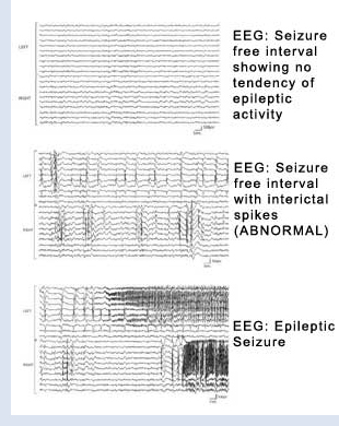
The electroencephalogram, also called the EEG, or the brain wave test was invented in 1929 and is therefore one of Neurologys oldest tests. Although it was once used to help diagnose a wide variety of neurologic problems, the EEG, like the lumbar puncture, has come to have a fairly limited usefulness. This usefulness is primarily in helping neurologists determine the cause of events commonly referred to as spells. Neurologic problems come in a variety of forms, and one of the more common forms is that of a transient and involuntary alteration in body position, thought content, or consciousness. In other words, spells. The electroencephalogram may help diagnose different kinds of seizure problems (epilepsy), feinting disorders (syncope), or sleep disorders (narcolepsy).
 An EEG is easy to perform, takes thirty or so minutes to do, and presents no risk to the patient. Electrodes are applied to the scalp and attached by long wires to the EEG machine, which today is a small computer. In preparation for the test the patient is asked to shampoo his or her hair but to avoid the use of additional oils, creams, lotions, gels, or other hair products. This test may be performed with no other special preparation or may be done with the patient in a sleep deprived state. Prior to a sleep deprived EEG the patient is asked to not sleep for a portion of the night so that he or she will be tired during the test. The reason for this is that fatigue lowers the brains defenses against displaying EEG abnormalities and thus makes it more likely that a diagnostic event will occur during the test. Also, at some point during an EEG the patient will usually be asked to hyperventilate for three minutes and at another time the patient will be subjected to what is called photic stimulation. During photic stimulation bright lights are flashed at different frequencies in front of the patients closed eyes. Like sleep deprivation, these procedures may help bring out EEG abnormalities that would not otherwise be visible.
The EEG is frequently a helpful test but it is not a perfect test, so that its results must be interpreted with a degree of caution. It is a 20 to 30 minute sample of the brains electrical activity and as such may not capture the abnormality that the doctor was looking for. This means that in order to come to an intelligent decision about your problem the EEG must be interpreted along with what the neurologist already knows about you from your own words and what any other observers have told the doctor about your problem. Routine EEGs are performed in neurology offices and hospitals, but occasionally it is necessary to refer a patient to a more specialized electroencephalography lab capable of performing prolonged brain wave recordings, called telemetry.
